Cellular ATP levels do not reflect mitochondrial defects associated with Barth syndrome
DOI:
https://doi.org/10.26124/bec.2025-0008Keywords:
adenosine triphosphate, adenosine diphosphate, mitochondria, cardiolipin, bioenergetics, Barth syndrome, adenosine monophosphateAbstract
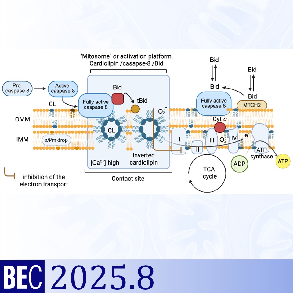
Barth syndrome (BTHS) is a rare X-linked genetic disease characterized by cardioskeletal myopathy, growth retardation, neutropenia, and abnormally high urinary 3-methylglutaconic acid. BTHS is known to result mutations in the TAFAZZIN gene that encodes an enzyme that is essential for the biosynthesis of mature cardiolipin, an inner membrane phosholipid required for several aspects of mitochondrial function. While defects in mitochondrial structure and metabolism have been widely reported in patient cells and TAFAZZIN-deficient animal models, levels of adenosine triphosphate (ATP) have been consistently found to be unchanged. This highlights the importance of evaluating deficiencies in mitochondrial function by multiple methods that are capable of capturing the many complex functions in regulating cellular metabolism, bioenergetics, ion and redox homeostasis and cell survival. Determining the mitochondrial defects responsible for BTHS has proved challenging given the development of profound structural and functional changes that can affect a wide variety of celllar processes. For this reason, future BTHS research should focus on the impacts of non-matured cardiolipin and mono-lysocardiolipin in mitochondrial membranes and their interactions rather than solely on cellular bioenergetics or ATP perturbations. Comprehensive evaluation of bioenergetic status that includes accurate measurements of metabolic flux (e.g., respirometry with specific substrates), levels of multiple high energy phosphates (ATP, ADP, AMP, phosphocreatine), along with NADPH/NADP+ and NADH/NAD+ ratios will help to better integrate and understand how mitochodrial bioenergetic contributes to the pathogenesis of BTHS and may be modified to reveal new therapeutic strategies.
Cite:
Petit PX (2025) Cellular ATP levels do not reflect mitochondrial defects associated with Barth syndrome. Bioenerg Commun 2025.8. https://doi.org/10.26124/bec.2025-0008
References
Acehan D, Vaz F, Houtkooper RH, James J, Moore V, Tokunaga C, Khuchua Z (2011) Cardiac and skeletal muscle defects in a mouse model of human Barth syndrome. https://doi.org/10.1074/jbc.M110.171439
Acehan D, Xu Y, Stokes DL, Schlame M (2007) Comparison of lymphoblast mitochondria from normal subjects and patients with Barth syndrome using electron microscopic tomography. https://doi.org/10.1038/labinvest.3700480
Ban T, Heymann JA, Song Z, Hinshaw JE, Chan DC (2010) OPA1 disease alleles causing dominant optic atrophy have defects in cardiolipin-stimulated GTP hydrolysis and membrane tubulation. https://doi.org/10.1093/hmg/ddq088
Barth PG, Van ’t Veer-Korthof ET, Van Delden L, Van Dam K, Van der Harten JJ, Kuipers JRG (1981) An X-linked mitochondrial disease affecting cardiac muscle, skeletal muscle and neutrophil leucocytes. Preliminary communication: Mefar B.V., Beetsterzwaag, The Netherlands.
Barth PG, Scholte HR, Berden JA, Van der Klei-Van Moorsel JM, Luyt-Houwen IE, Van 't Veer-Korthof ET, Van der Harten JJ, Sobotka-Plojhar MA (1983) An X-linked mitochondrial disease affecting cardiac muscle, skeletal muscle and neutrophil leucocytes. https://doi.org/10.1016/0022-510X(83)90209-5
Barth PG, Valianpour F, Bowen VM, Lam J, Duran M, Vaz FM, Wanders RJ (2004) X-linked cardioskeletal myopathy and neutropenia (Barth syndrome): an update. https://doi.org/10.1002/ajmg.a.20660
Bazan S, Mileykovskaya E, Mallampalli VK, Heacock P, Sparagna GC, Dowhan W (2013) Cardiolipin-dependent reconstitution of respiratory supercomplexes from purified Saccharomyces cerevisiae complexes III and IV. https://doi.org/10.1074/jbc.M112.425876
Brand MD, Nicholls DG (2011) Assessing mitochondrial dysfunction in cells. https://doi.org/10.1042/BJ20110162
Brandner K, Mick DU, Frazier AE, Taylor RD, Meisinger C, Rehling P (2005) Taz1, an outer mitochondrial membrane protein, affects stability and assembly of inner membrane protein complexes: implications for Barth Syndrome. https://doi.org/10.1091/mbc.e05-03-0256
Brault JJ, Conway SJ (2025) What can ATP content tell us about Barth syndrome muscle phenotypes? https://doi.org/10.20517/jtgg.2024.83
Cardoso LHD, Doerrier C, Gnaiger E (2021) Magnesium Green for fluorometric measurement of ATP production does not interfere with mitochondrial respiration. https://doi.org/10.26124/bec:2021-0001
Chatham JC, Young ME (2012) Metabolic remodeling in the hypertrophic heart: fuel for thought. https://doi.org/10.1161/CIRCRESAHA.112.277392
Chu CT, Bayir H, Kagan VE (2014) LC3 binds externalized cardiolipin on injured mitochondria to signal mitophagy in neurons: implications for Parkinson disease. https://doi.org/10.4161/auto.27191
de Kok MJC, Schaapherder AF, Wust RCI, Zuiderwijk M, Bakker JA, Lindeman JHN, Le Devedec SE (2021) Circumventing the Crabtree effect in cell culture: A systematic review. https://doi.org/10.1016/j.mito.2021.03.014
de Taffin de Tilques M, Lasserre JP, Godard F, Sardin E, Bouhier M, Le Guedard M, Kucharczyk R, Petit PX, Testet E, di Rago JP, Tribouillard-Tanvier D (2018) Decreasing cytosolic translation is beneficial to yeast and human Tafazzin-deficient cells. https://doi.org/10.15698/mic2018.05.629
Desousa BR, Kim KK, Jones AE, Ball AB, Hsieh WY, Swain P, Morrow DH, Brownstein AJ, Ferrick DA, Shirihai OS, Neilson A, Nathanson DA, Rogers GW, Dranka BP, Murphy AN, Affourtit C, Bensinger SJ, Stiles L, Romero N, Divakaruni AS (2023) Calculation of ATP production rates using the Seahorse XF Analyzer. https://doi.org/10.15252/embr.202256380
DeVay RM, Dominguez-Ramirez L, Lackner LL, Hoppins S, Stahlberg H, Nunnari J (2009) Coassembly of Mgm1 isoforms requires cardiolipin and mediates mitochondrial inner membrane fusion. https://doi.org/10.1083/jcb.200906098
Divakaruni AS, Jastroch M (2022) A practical guide for the analysis, standardization and interpretation of oxygen consumption measurements. https://doi.org/10.1038/s42255-022-00619-4
Doerrier C, Garcia-Souza LF, Krumschnabel G, Wohlfarter Y, Meszaros AT, Gnaiger E (2018) High-Resolution FluoRespirometry and OXPHOS Protocols for Human Cells, Permeabilized Fibers from Small Biopsies of Muscle, and Isolated Mitochondria. https://doi.org/10.1007/978-1-4939-7831-1_3
Dudek J, Maack C (2017) Barth syndrome cardiomyopathy. https://doi.org/10.1093/cvr/cvx014
Gawrisch K (2012) Tafazzin senses curvature. https://doi.org/10.1038/nchembio.1068
Gebert N, Joshi AS, Kutik S, Becker T, McKenzie M, Guan XL, Mooga VP, Stroud DA, Kulkarni G, Wenk MR, Rehling P, Meisinger C, Ryan MT, Wiedemann N, Greenberg ML, Pfanner N (2009) Mitochondrial cardiolipin involved in outer-membrane protein biogenesis: implications for Barth syndrome. https://doi.org/10.1016/j.cub.2009.10.074
Gendron MC, Schrantz N, Métivier D, Kroemer G, Maciorowska Z, Sureau F, Koester S, Petit PX (2001) Oxidation of pyridine nucleotides during Fas- and ceramide-induced apoptosis in Jurkat cells: correlation with changes in mitochondria, glutathione depletion, intracellular acidification and caspase 3 activation. https://pmc.ncbi.nlm.nih.gov/articles/PMC1221579/
Gonzalvez F, D'Aurelio M, Boutant M, Moustapha A, Puech JP, Landes T, Arnauné-Pelloquin L, Vial G, Taleux N, Slomianny C, Wanders RJ, Houtkooper RH, Bellenguer P, Møller IM, Gottlieb E, Vaz FM, Manfredi G, Petit PX (2013) Barth syndrome: cellular compensation of mitochondrial dysfunction and apoptosis inhibition due to changes in cardiolipin remodeling linked to tafazzin (TAZ) gene mutation. https://doi.org/10.1016/j.bbadis.2013.03.005
Gonzalvez F, Pariselli F, Dupaigne P, Budihardjo I, Lutter M, Antonsson B, Diolez P, Manon S, Martinou JC, Goubern M, Wang X, Bernard S, Petit PX (2005) tBid interaction with cardiolipin primarily orchestrates mitochondrial dysfunctions and subsequently activates Bax and Bak. https://doi.org/10.1038/sj.cdd.4401571
Gonzalvez F, Schug ZT, Houtkooper RH, MacKenzie ED, Brooks DG, Wanders RJ, Petit PX, Vaz FM, Gottlieb E (2008) Cardiolipin provides an essential activating platform for caspase-8 on mitochondria. https://doi.org/10.1083/jcb.200803129
Gu Z, Valianpour F, Chen S, Vaz FM, Hakkaart GA, Wanders RJ, Greenberg ML (2004) Aberrant cardiolipin metabolism in the yeast taz1 mutant: a model for Barth syndrome. https://doi.org/10.1046/j.1365-2958.2003.03802.x
Gu X, Ma Y, Liu Y, Wan Q (2021) Measurement of mitochondrial respiration in adherent cells by Seahorse XF96 Cell Mito Stress Test. https://doi.org/10.1016/j.xpro.2020.100245
Hancock CR, Brault JJ, Wiseman RW, Terjung RL, Meyer RA (2005) 31P-NMR observation of free ADP during fatiguing, repetitive contractions of murine skeletal muscle lacking AK1. https://doi.org/10.1152/ajpcell.00621.2004
Handel ME, Brand MD, Mookerjee SA (2019) The Whys and Hows of Calculating Total Cellular ATP Production Rate. https://doi.org/10.1016/j.tem.2019.04.007
He Q (2010) Tafazzin knockdown causes hypertrophy of neonatal ventricular myocytes. https://doi.org/10.1152/ajpheart.00098.2010
Heit B, Yeung T, Grinstein S (2011) Changes in mitochondrial surface charge mediate recruitment of signaling molecules during apoptosis. https://doi.org/10.1152/ajpcell.00139.2010
Ikon N, Su B, Hsu FF, Forte TM, Ryan RO (2015) Exogenous cardiolipin localizes to mitochondria and prevents TAZ knockdown-induced apoptosis in myeloid progenitor cells. https://doi.org/10.1016/j.bbrc.2015.07.012
Jiang F, Ryan MT, Schlame M, Zhao M, Gu Z, Klingenberg M, Pfanner N, Greenberg ML. (2000) Absence of cardiolipin in the crd1 null mutant results in decreased mitochondrial membrane potential and reduced mitochondrial function. https://doi.org/10.1074/jbc.M909868199
Joshi AS, Thompson MN, Fei N, Huttemann M, Greenberg ML (2012) Cardiolipin and mitochondrial phosphatidylethanolamine have overlapping functions in mitochondrial fusion in Saccharomyces cerevisiae. https://doi.org/10.1074/jbc.M111.330167
Kadenbach B, Mende P, Kolbe HV, Stipani I, Palmieri F (1982) The mitochondrial phosphate carrier has an essential requirement for cardiolipin. https://doi.org/10.1016/0014-5793(82)80498-5
Khuchua Z, Yue Z, Batts L, Strauss AW (2006) A zebrafish model of human Barth syndrome reveals the essential role of tafazzin in cardiac development and function. https://doi.org/10.1161/01.RES.0000233378.95325.ce
Kim TH, Zhao Y, Ding WX, Shin JN, He X, Seo YW, Chen J, Rabinowich H, Amoscato AA, Yin XM (2004) Bid-cardiolipin interaction at mitochondrial contact site contributes to mitochondrial cristae reorganization and cytochrome C release. https://doi.org/10.1091/mbc.e03-12-0864
Koshkin V, Greenberg ML (2000) Oxidative phosphorylation in cardiolipin-lacking yeast mitochondria. https://pubmed.ncbi.nlm.nih.gov/10769171/
Law AS, Hafen PS, Brault JJ (2022) Liquid chromatography method for simultaneous quantification of ATP and its degradation products compatible with both UV-Vis and mass spectrometry. https://doi.org/10.1016/j.jchromb.2022.123351
Li XX, Tsoi B, Li YF, Kurihara H, He RR (2015) Cardiolipin and its different properties in mitophagy and apoptosis. https://doi.org/10.1369/0022155415574818
Manganelli V, Capozzi A, Recalchi S, Signore M, Mattei V, Garofalo T, Misasi R, Degli Esposti M, Sorice M (2015) Altered Traffic of Cardiolipin during Apoptosis: Exposure on the Cell Surface as a Trigger for "Antiphospholipid Antibodies". https://doi.org/10.1155/2015/847985
Marvin JS, Kokotos AC, Kumar M, Pulido C, Tkachuk AN, Yao JS, Brown TA, Ryan TA (2024) iATPSnFR2: A high-dynamic-range fluorescent sensor for monitoring intracellular ATP. https://doi.org/10.1073/pnas.2314604121
McKenzie M, Lazarou M, Thorburn DR, Ryan MT (2006) Mitochondrial respiratory chain supercomplexes are destabilized in Barth Syndrome patients. https://doi.org/10.1016/j.jmb.2006.06.057
Miller SG, Hafen PS, Brault JJ (2019) Increased Adenine Nucleotide Degradation in Skeletal Muscle Atrophy. https://doi.org/10.3390/ijms21010088
Mookerjee SA, Brand MD (2015) Measurement and Analysis of Extracellular Acid Production to Determine Glycolytic Rate. https://doi.org/10.3791/53464
Mookerjee SA, Gerencser AA, Nicholls DG, Brand MD (2017) Quantifying intracellular rates of glycolytic and oxidative ATP production and consumption using extracellular flux measurements. https://doi.org/10.1074/jbc.M116.774471
Mookerjee SA, Goncalves RLS, Gerencser AA, Nicholls DG, Brand MD (2015) The contributions of respiration and glycolysis to extracellular acid production. https://doi.org/10.1016/j.bbabio.2014.10.005
Neustein HB, Lurie PR, Dahms B, Takahashi M (1979) An X-linked recessive cardiomyopathy with abnormal mitochondria. https://doi.org/10.1542/peds.64.1.24
Nickel A, Loffler J, Maack C (2013) Myocardial energetics in heart failure. https://doi.org/10.1007/s00395-013-0358-9
Noel H, Pande SV (1986) An essential requirement of cardiolipin for mitochondrial carnitine acylcarnitine translocase activity. Lipid requirement of carnitine acylcarnitine translocase. https://doi.org/10.1111/j.1432-1033.1986.tb09463.x
Patil VA, Greenberg ML (2013) Cardiolipin-mediated cellular signaling. https://doi.org/10.1007/978-94-007-6331-9_11
Pelletier M, Billingham LK, Ramaswamy M, Siegel RM (2014) Extracellular flux analysis to monitor glycolytic rates and mitochondrial oxygen consumption. https://doi.org/10.1016/B978-0-12-416618-9.00007-8
Pfeiffer K, Gohil V, Stuart RA, Hunte C, Brandt U, Greenberg ML, Schagger H (2003) Cardiolipin stabilizes respiratory chain supercomplexes. https://doi.org/10.1074/jbc.M308366200
Phoon CK, Acehan D, Schlame M, Stokes DL, Edelman-Novemsky I, Yu D, Xu Y, Viswanathan N, Ren M (2012) Tafazzin knockdown in mice leads to a developmental cardiomyopathy with early diastolic dysfunction preceding myocardial noncompaction. https://doi.org/10.1161/JAHA.111.000455
Robinson NC (1993) Functional binding of cardiolipin to cytochrome c oxidase. https://doi.org/10.1007/BF00762857
Saini-Chohan HK, Holmes MG, Chicco AJ, Taylor WA, Moore RL, McCune SA, Hickson-Bick DL, Hatch GM, Sparagna GC (2009) Cardiolipin biosynthesis and remodeling enzymes are altered during development of heart failure. https://doi.org/10.1194/jlr.M800561-JLR200
Saric A, Andreau K, Armand AS, Moller IM, Petit PX (2015) Barth Syndrome: From Mitochondrial Dysfunctions Associated with Aberrant Production of Reactive Oxygen Species to Pluripotent Stem Cell Studies. https://doi.org/10.3389/fgene.2015.00359
Schlame M, Rua D, Greenberg ML (2000) The biosynthesis and functional role of cardiolipin. https://doi.org/10.1016/s0163-7827(00)00005-9
Stanley PE, Williams SG (1969) Use of the liquid scintillation spectrometer for determining adenosine triphosphate by the luciferase enzyme. https://doi.org/10.1016/0003-2697(69)90323-6
Tullson PC, Terjung RL (1990) Adenine nucleotide degradation in striated muscle. https://doi.org/10.1055/s-2007-1024854
Tullson PC, Whitlock DM, Terjung RL (1990) Adenine nucleotide degradation in slow-twitch red muscle. https://doi.org/10.1152/ajpcell.1990.258.2.C258
Vaz FM, Houtkooper RH, Valianpour F, Barth PG, Wanders RJ (2003) Only one splice variant of the human TAZ gene encodes a functional protein with a role in cardiolipin metabolism. https://doi.org/10.1074/jbc.M305956200
Wang G, McCain ML, Yang L, He A, Pasqualini FS, Agarwal A, Yuan H, Jiang D, Zhang D, Zangi L, Geva J, Roberts AE, Ma Q, Ding J, Chen J, Wang DZ, Li K, Wang J, Wanders RJ, Kulik W, Vaz FM, Laflamme MA, Murry CE, Chien KR, Kelley RI, Church GM, Parker KK, Pu WT (2014) Modeling the mitochondrial cardiomyopathy of Barth syndrome with induced pluripotent stem cell and heart-on-chip technologies. https://doi.org/10.1038/nm.3545
Wang X, Chen XJ (2015) A cytosolic network suppressing mitochondria-mediated proteostatic stress and cell death. https://doi.org/10.1038/nature14859
Xu Y, Kelley RI, Blanck TJ, Schlame M (2003) Remodeling of cardiolipin by phospholipid transacylation. https://doi.org/10.1074/jbc.M307382200
Xu Y, Sutachan JJ, Plesken H, Kelley RI, Schlame M (2005) Characterization of lymphoblast mitochondria from patients with Barth syndrome. https://doi.org/10.1038/labinvest.3700274
Xu Y, Condell M, Plesken H, Edelman-Novemsky I, Ma J, Ren M, Schlame M (2006) A Drosophila model of Barth syndrome. https://doi.org/10.1073/pnas.0603242103
Xu Y, Malhotra A, Ren M, Schlame M (2006) The enzymatic function of tafazzin. https://doi.org/10.1074/jbc.M606100200
Yang NC, Ho WM, Chen YH, Hu ML (2002) A convenient one-step extraction of cellular ATP using boiling water for the luciferin-luciferase assay of ATP. https://doi.org/10.1006/abio.2002.5698
Yurista SR, Nguyen CT, Rosenzweig A, de Boer RA, Westenbrink BD (2021) Ketone bodies for the failing heart: fuels that can fix the engine? https://doi.org/10.1016/j.tem.2021.07.006
Zhang M, Mileykovskaya E, Dowhan W (2002) Gluing the respiratory chain together. Cardiolipin is required for supercomplex formation in the inner mitochondrial membrane. https://doi.org/10.1074/jbc.C200551200
Zhu S, Chen Z, Zhu M, Shen Y, Leon LJ, Chi L, Spinozzi S, Tan C, Gu Y, Nguyen A, Zhou Y, Feng W, Vaz FM, Wang X, Gustafsson AB, Evans SM, Kunfu O, Fang X (2021) Cardiolipin Remodeling Defects Impair Mitochondrial Architecture and Function in a Murine Model of Barth Syndrome Cardiomyopathy. https://doi.org/10.1161/CIRCHEARTFAILURE.121.008289
Downloads
Published
License
Copyright (c) 2025 Dr. Patrice X Petit

This work is licensed under a Creative Commons Attribution-NonCommercial-NoDerivatives 4.0 International License.